
Surface Architectures for the Manipulation of Cell Adhesion
Controlling the reaction of cells to surface properties is important in many regards. We are engaged in projects that combine surface topography and surface chemistry as well as mechanical properties of the used materials to manipulate cells and cell organells in different ways.
In one project we use micropillar arrays in which the top surfaces of the pillars and the areas in between the pillars are made from materials that are either cell attractive of cell repellent. Cells react to this environment in different ways. Depending on the actual feature size some cells literally wrap around the pillars in a way that also leads to a strong deformation of the cell's nucleus. Our example in Figure 1 shows such a situation. The nucleus of these bone cancer sells is stained in blue and the pillars of this top view are displayed in red.
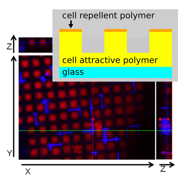
Figure 1: Microscopic image of bone cancer cells on micropillar arrays: The nuclei are strongly deformed as the cell reacts to the microstructure and surface chemistry. The pillar tops are cell repellent the spaces in between are cell attractive.
In a related project we study the behavior of human cardiomyocytes (CM) on similar pillar arrays. Only the tops of the pillars are cell attractive and the cells are located only there. The substrates induce cell alignment and a sarcomeric organization in the cells resembling closely that of cells under physiological conditions. During contraction of the cells the bending of elastic pillars can be followed optically and the force exerted by the cell onto the substrate during contraction can be determined and compared to the calcium transient to find a correlation between cell physiology and cell mechanics.
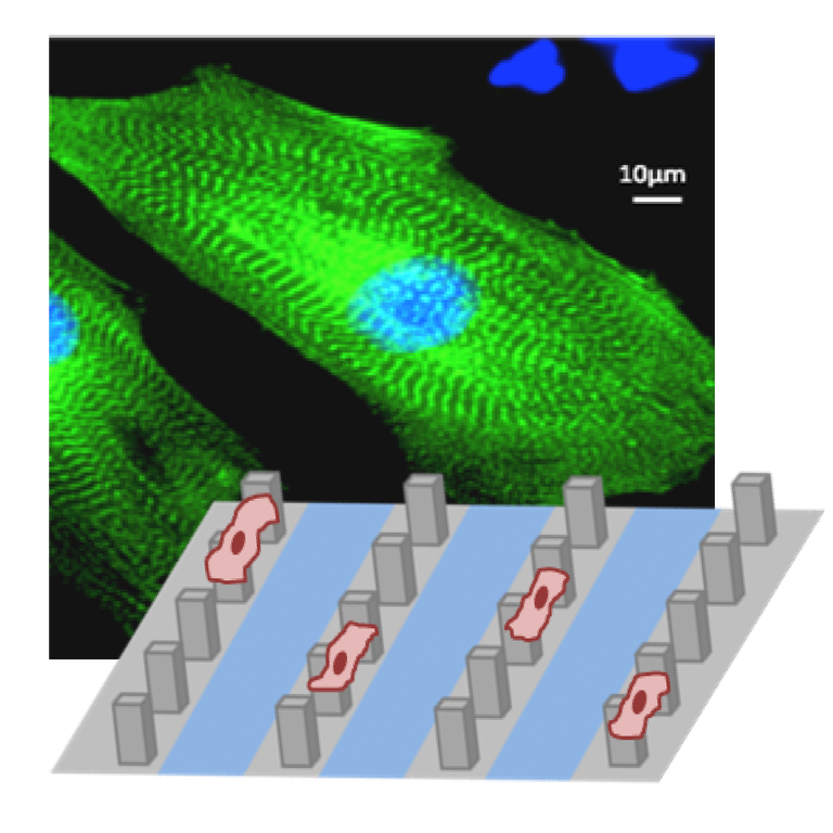
Figure 2: Microscopic image of cardiomyocytes on micropillar arrays.